Rumored Buzz on Babyecho
Table of ContentsWhat Does Babyecho Mean?The 6-Second Trick For BabyechoBabyecho for BeginnersSome Ideas on Babyecho You Need To KnowMore About BabyechoSome Of BabyechoEverything about Babyecho

A c-section is surgical procedure in which your baby is born through a cut that your doctor makes in your belly and uterus. No matter what an ultrasound reveals, speak to your carrier concerning the very best take care of you and your child - fetal doppler at home. Last evaluated: October, 2019
During this scan, they will certainly check the baby is expanding in the right area, whether there is greater than one infant and they will additionally examine your baby's advancement thus far. This screening is available in between 10 14 weeks of maternity and is made use of to evaluate the chances of your child being birthed with one or even more of these conditions.
Not known Incorrect Statements About Babyecho
It entails a combined examination of an ultrasound check and a blood examination. Throughout the check, the sonographer will certainly measure the liquid at the back of the child's neck to identify 'nuchal clarity' - https://lwccareers.lindsey.edu/profiles/4681929-leroy-parker. They will certainly after that compute the opportunity of your child having Down's, Edwards' or Patau's syndrome using your age, the blood test and scan outcomes
During this scan, the sonographer look for architectural and developing problems in the baby. Throughout this scan consultation, you might be offered testings for HIV, syphilis and liver disease B by a specialist midwife. Sometimes, a third-trimester scan is recommended by your midwife adhering to the results of previous examinations, previous issues or existing medical problems.
The standard 2D ultrasound creates level and detailed photos which can be used to see your child's inner organs and help identify any type of internal issues. These black and white photos help the sonographer establish the child's gestation, growth, heartbeat, development and size. Some expectant mommies select to have a 3D ultrasound check due to the fact that they reveal even more of a real-life photo of the infant.
How Babyecho can Save You Time, Stress, and Money.
3D ultrasound scans reveal still photos of your child's external body instead of their withins, so you can see the form of the child's face attributes. 4D ultrasound scans are similar to 3D scans yet they show a relocating video instead of still photos. This records highlights and shadows much better, as a result creating a more clear photo of the child's face and activities.

or (8-11 weeks) (11-14 weeks) (14-18 weeks) (19-23 weeks) or (24-42 weeks) Suggested at Optional -, a lot more often in some problems This scan is done to and to figure out an (EDD). A is discovered during this scan. Many moms and dads go with this scan for. Is vital prior to the blood test called as (NIPT) to determine the.
How Babyecho can Save You Time, Stress, and Money.
Occasionally a may be called for to get and a clearer image. This is generally carried out and periodically a might be required. Verify that the infant's heart is existing; To much more precisely. This might not be essential in, where the from the is much more precise; To; To diagnose whether and to examine whether there is sharing of placenta, which will require close surveillance in maternity; To analyze the consisting of measurement of; To see if there is a reduced or high opportunity for the infant to be impacted with such as Down's Disorder, Edward's Syndrome and; If any type of, better relating to will certainly be provided at the very same assessment by myself.
Please see below. It coincides as 19-22 weeks, but some may be or in the and it may to. Normally this is provided if there are such as spina bifida or if parents are eager to understand the earlier. These scans might be done, nevertheless several of the and therefore, a is needed to This scan is done typically at.
7 Simple Techniques For Babyecho

In addition, the can be by by an. () The method nearer the is helpful to. Sometimes, an which was before may be.
Babyecho for Beginners
If, these scans may be to. on the searchings for, a may be used. Throughout all the, a 3D scan (of the child) can likewise be performed. The hinges on the position of the,,, amount of and. This consists of, together with; This includes, along with (14-20 weeks).
A check is crucial prior to this examination is done. If you're looking for, arrange an examination with Dr Sankaran through her. Obstetrics & gynaecology in London.
How Babyecho can Save You Time, Stress, and Money.
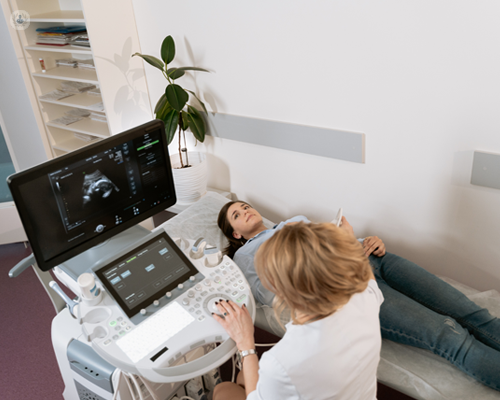
A prenatal ultrasound check is a diagnostic strategy that utilizes high-frequency audio waves to develop a photo of your unborn child. Ultrasounds may be carried out at different times throughout pregnancy for various reasons. The examination can offer important information, aiding women and their health-care service providers manage and care for the maternity and the fetus.
A transducer is put into the vagina and relaxes against the rear of the vaginal useful source canal to create a photo. A transvaginal ultrasound creates a sharper picture and is typically used in very early pregnancy. Ultrasound makers are about the size of a grocery cart. A television display for seeing the images is affixed to the device (https://pagespeed.web.dev/analysis/https-babyfetaldoppler-com/wlbdlhbwfi?form_factor=mobile).